
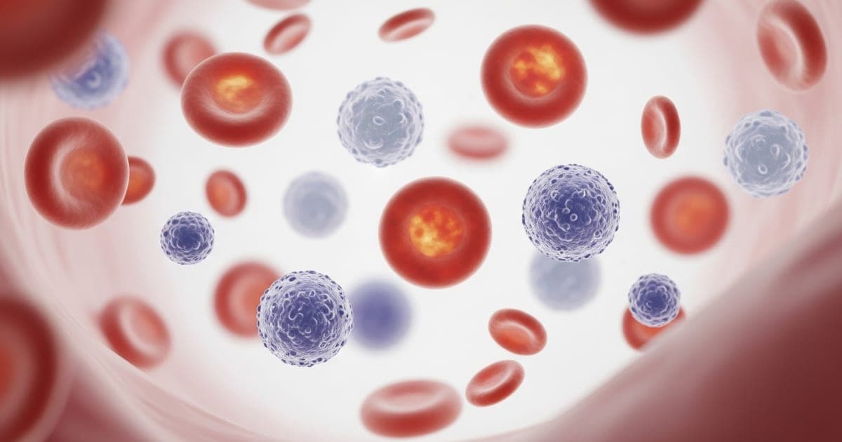
NRBCs (nucleated red blood cells) are immature red blood cell precursors that belong in the bone marrow, not in circulating blood. Finding even one NRBC per 100 white blood cells on an adult CBC is flagged as abnormal. Their presence signals that something is forcing the marrow to release cells before they finish maturing — and identifying that cause becomes the clinical priority.
What Is an NRBC? The Maturation Sequence
Red blood cells develop through six stages in the bone marrow:
- Proerythroblast — large, nucleus-dominant precursor
- Basophilic erythroblast — begins hemoglobin synthesis
- Polychromatic erythroblast — hemoglobin accumulation continues
- Orthochromatic erythroblast (NRBC) — last stage with intact nucleus; recognizable on a blood smear
- Reticulocyte — nucleus expelled; released into circulation
- Mature RBC — anucleate, biconcave disc, fully functional
At stage 4, the bone marrow normally holds these cells until the nucleus is expelled. If the marrow is overwhelmed, or if the spleen (which filters escaped NRBCs) is absent or non-functional, NRBCs enter circulation prematurely.
Normal NRBC Values
Adults: zero. Any NRBC detected is abnormal and will trigger a pathology review.
Newborns (0–7 days): NRBCs are physiologically normal in the first days of life as the newborn's marrow adjusts. They normalize within the first week and are absent in healthy infants after one month.
What Causes NRBCs in Adults?
| Mechanism | Specific Cause | What's Driving It |
|---|---|---|
| Stress erythropoiesis (marrow in overdrive) | Severe hemolytic anemia (autoimmune, sickle cell crisis, G6PD deficiency) | RBCs destroyed faster than marrow can replace; immature cells released early |
| Severe acute blood loss / hemorrhage | EPO surge drives emergency production; NRBCs released prematurely | |
| Severe hypoxia (respiratory failure, CO poisoning, high-altitude) | Low oxygen → high EPO → accelerated marrow output | |
| Severe iron deficiency anemia | Marrow dysfunction from chronic iron starvation | |
| Bone marrow infiltration | Metastatic cancer (breast, prostate, lung most common) | Tumor cells displace normal marrow → leukoerythroblastic reaction |
| Myelofibrosis | Fibrotic marrow replacement forces extramedullary hematopoiesis | |
| Leukemia / lymphoma | Malignant infiltration displaces erythroid precursors | |
| Splenic dysfunction | Asplenia (surgical or functional — sickle cell) | NRBC filter absent; even low-level releases reach circulation |
| Hypersplenism / severe splenic overload | Overwhelmed spleen fails to clear all NRBCs |
How NRBCs Are Reported on a CBC
Modern automated analyzers detect NRBCs and report them as NRBC% (per 100 WBCs). One important technical detail: analyzers initially count NRBCs as white blood cells because they detect the nucleus. This inflates the WBC. When NRBCs are flagged, the lab corrects the count:
Corrected WBC = Uncorrected WBC × [100 ÷ (100 + NRBC count)]
Example: reported WBC 12,000 with 20 NRBCs/100 WBCs → corrected WBC = 12,000 × (100 ÷ 120) = 10,000. This distinction matters — an apparently elevated WBC can normalize once NRBCs are accounted for.
NRBCs as a Prognostic Marker in Critical Illness
In ICU settings, NRBC presence is an independent predictor of 30-day mortality, even after adjusting for other severity scores. Multiple studies show that critically ill patients with detectable NRBCs have significantly higher mortality than those without. Rising NRBC counts during ICU admission correlate with worsening physiological stress. In sepsis, major trauma, and multi-organ failure, NRBC monitoring adds prognostic information beyond standard labs.
What Happens After NRBCs Are Found?
Finding NRBCs on a CBC triggers a structured workup:
- Peripheral blood smear — a hematologist reviews for NRBC confirmation and co-existing abnormalities (tear-drop cells, blast forms, hypersegmented neutrophils)
- Reticulocyte count — elevated = stress erythropoiesis (marrow responding); low = marrow suppression or infiltration
- Hemolysis markers — LDH, total bilirubin, haptoglobin to identify hemolytic causes
- Direct antiglobulin test (DAT) — positive result indicates autoimmune hemolytic anemia
- Bone marrow biopsy — required if infiltration, myelofibrosis, or hematologic malignancy is suspected
- Imaging — CT or MRI to assess for splenomegaly, metastatic disease, or lymphoma
Getting a CBC with Differential at Home
A CBC with differential — the test that detects and reports NRBCs — can be ordered and collected through mobile phlebotomy. A licensed phlebotomist comes to your home, collects the sample, and it is processed at the same certified reference lab your doctor uses. If you need a provider to issue the requisition, a telehealth consultation can be coordinated before the draw.
NRBCs in specific high-risk conditions
While NRBCs can appear in any condition that severely stresses the bone marrow, certain diagnoses are particularly associated with them and carry specific clinical implications:
Asplenia (absent or non-functional spleen): The spleen is the primary filter for NRBCs in adults. Patients who have had their spleen removed (splenectomy) or who have functional asplenia from sickle cell disease, celiac disease, or certain autoimmune conditions will have persistent low-level NRBCs on their CBC — this is expected and benign in this context. It does not trigger the same workup as NRBCs in a patient with a functioning spleen.
Sickle cell crisis: During a vaso-occlusive crisis, hypoxia and accelerated RBC destruction drive a surge in erythropoietin. The marrow releases reticulocytes and sometimes NRBCs as it tries to compensate. NRBC counts in sickle cell crisis often normalize between episodes.
Myelophthisic anemia: When bone marrow is replaced or infiltrated by abnormal cells — metastatic cancer, myelofibrosis, lymphoma, granulomas — the normal marrow architecture is disrupted and immature cells including NRBCs spill into the bloodstream. This pattern (called leukoerythroblastosis) — NRBCs plus immature white cells (band neutrophils, metamyelocytes) plus large abnormal platelets — is a red flag for bone marrow infiltration and almost always prompts a bone marrow biopsy.
Sepsis and critical illness: NRBCs in ICU patients are an independent predictor of mortality. In this setting, they likely reflect a combination of hypoxic stress on the marrow and breakdown of normal marrow retention mechanisms. Their presence should prompt re-evaluation of the severity of illness, not be dismissed as incidental.
The diagnostic workup after NRBCs are detected
Finding NRBCs on an adult CBC initiates a structured evaluation. The urgency and depth depend on the clinical context:
- Peripheral blood smear review — a laboratory technologist or hematopathologist manually examines the smear. This confirms the NRBC count, evaluates cell morphology (teardrop cells suggest myelofibrosis; sickle cells confirm SCD; hypersegmented neutrophils suggest megaloblastic anemia), and checks for co-occurring abnormalities in white cells and platelets.
- Complete history and physical — splenectomy history, malignancy history, recent acute illness, family history of hemoglobinopathy, medication review.
- Directed laboratory workup based on the most likely cause:
- Hemolytic workup: LDH, haptoglobin, direct Coombs test, bilirubin
- Iron and nutritional studies: ferritin, B12, folate
- Hemoglobin electrophoresis if thalassemia or sickle cell is suspected
- Inflammatory markers: CRP, ESR, procalcitonin
- Imaging: CT chest/abdomen/pelvis if malignancy or marrow infiltration is suspected
- Bone marrow biopsy — indicated when malignancy, myelofibrosis, aplastic anemia, or unexplained persistent NRBCs are the leading possibilities after initial workup. This is typically ordered by a hematologist.
NRBCs alone do not point to a single diagnosis. They are a signal — the specificity lies in the combination of smear findings, clinical context, and supporting labs. Most patients with NRBCs will have a clear cause identified within the first round of targeted testing.
Sources and further reading
CBC With Differential — Drawn at Home
An NRBC result comes from a standard CBC with differential draw. A certified mobile phlebotomist comes to you — the sample goes to your provider's certified reference lab.
Book a visit →